Sunday 22 October 2023

Physiotherapist Meaning in Gujarati


Physiotherapist Meaning in Gujarati

ફિઝિયોથેરાપિસ્ટ એ એક આરોગ્ય સંભાળ વ્યવસાયી છે જે શારીરિક સારવારનો ઉપયોગ કરીને લોકોને તેમની ગતિશીલતા, કાર્યક્ષમતા અને જીવનની ગુણવત્તા સુધારવામાં મદદ કરે છે. ફિઝિયોથેરાપિસ્ટ્સ વિવિધ પ્રકારની સ્થિતિઓની સારવાર કરે છે, જેમાં શામેલ છે:

  • સ્નાયુઓ અને સાંધામાં દુખાવો
  • ઈજાઓ
  • સ્ટ્રોક
  • મલ્ટીપલ સ્કલેરોસિસ
  • પાર્કિન્સન્સ રોગ
  • અસ્થિવા
  • કેન્સર

ફિઝિયોથેરાપીની સારવારમાં વ્યાયામ, મસાજ, હીટ થેરાપી, કોલ્ડ થેરાપી અને ઇલેક્ટ્રિકલ સ્ટીમ્યુલેશનનો સમાવેશ થઈ શકે છે. ફિઝિયોથેરાપિસ્ટ્સ દર્દીઓને તેમની સ્થિતિને સમજવામાં અને તેમની સ્વાસ્થ્ય સુધારવા માટે જીવનશૈલીમાં ફેરફાર કરવામાં મદદ કરે છે.

ફિઝિયોથેરાપિસ્ટ બનવા માટે, તમારે ફિઝિયોથેરાપીમાં સ્નાતકની ડિગ્રી પૂર્ણ કરવી આવશ્યક છે. ફિઝિયોથેરાપીના ક્ષેત્રમાં વિવિધ પ્રકારની વિશેષતાઓ છે, જેમ કે:

  • સ્પોર્ટ્સ ફિઝિયોથેરાપી
  • ન્યુરોલોજિકલ ફિઝિયોથેરાપી
  • કાર્ડિયોપલ્મોનરી ફિઝિયોથેરાપી
  • પેડિયાટ્રિક ફિઝિયોથેરાપી
  • ગેરિએટ્રિક ફિઝિયોથેરાપી

જો તમે ફિઝિયોથેરાપીમાં કારકિર્દી બનાવવામાં રસ ધરાવો છો, તો તમારે વિજ્ઞાન, ગણિત અને જીવવિજ્ઞાનમાં મજબૂત પ nền ધરાવવો જોઈએ. તમારે સારા સંચાર અને લોકો સાથે કામ કરવાની ક્ષમતા પણ હોવી જોઈએ.

Sunday 16 April 2023

Cervical Radiculopathy: Cause, Symptoms, Treatment, Exercise

What is a Cervical Radiculopathy?


Cervical Radiculopathy

Cervical radiculopathy refers to a condition where there is compression or irritation of one or more nerve roots in the cervical spine (neck region), leading to pain, numbness, or weakness in the areas of the body that the affected nerve supplies. 

The condition is often caused by degenerative changes in the spine, such as herniated discs, bone spurs, or narrowing of the spinal canal (stenosis). It can also be caused by trauma or injury to the neck, or by inflammation or infection in the area. Treatment options for cervical radiculopathy may include rest, physical therapy, medication, or in some cases, surgery.

Related Anatomy


The cervical spine, or neck region, consists of seven vertebrae, labeled C1 to C7, that are separated by intervertebral discs. Between each pair of vertebrae, a pair of nerve roots emerge from the spinal cord and exit the spine through small openings called foramina. These nerve roots then branch out to supply sensation and motor function to various parts of the body, such as the arms, shoulders, and hands.

The cervical spine also contains various ligaments, muscles, and other soft tissues that provide stability and support to the vertebrae and the surrounding structures. The ligaments help hold the vertebrae together, while the muscles allow for movement of the neck and head. The spinal cord, which is a long, thin bundle of nerve fibers that runs from the brain down through the spine, is protected by the vertebrae and surrounded by protective membranes called meninges.

Causes of Cervical Radiculopathy

Cervical radiculopathy can be caused by a variety of factors that put pressure on or irritate the nerve roots in the neck. Some common causes include:

  • Herniated or bulging discs: When a disc in the cervical spine ruptures or bulges out of its normal position, it can press against the nerve root, leading to pain and other symptoms.
  • Bone spurs: Bony growths that develop on the vertebrae due to age or degeneration can also impinge on the nerve roots, causing cervical radiculopathy.
  • Degenerative disc disease: As the discs in the spine naturally age and wear down, they can lose height and become less flexible, leading to pressure on the nerve roots.
  • Spinal stenosis: A narrowing of the spinal canal or foramina can occur due to conditions such as arthritis or degenerative disc disease, compressing the nerve roots.
  • Trauma or injury: A sudden injury or trauma to the neck, such as a whiplash injury, can damage the nerve roots and cause symptoms.
  • Infections or tumors: In rare cases, infections or tumors in the cervical spine can put pressure on the nerve roots and cause cervical radiculopathy.
  • Poor posture: Prolonged sitting or standing in a slouched position or any position that places stress on the neck can cause or exacerbate cervical radiculopathy.

Symptoms of Cervical Radiculopathy

The symptoms of cervical radiculopathy can vary depending on the location and severity of the nerve root compression or irritation. Some common symptoms may include:

  • Pain: Pain may be felt in the neck, shoulder, arm, or hand on the affected side. The pain may be sharp, burning, or dull, and may worsen with certain movements or positions.
  • Numbness or tingling: Numbness or tingling may be felt in the same areas as the pain, and may be accompanied by a pins-and-needles sensation.
  • Weakness: Weakness or difficulty with certain movements, such as lifting or gripping objects, may occur in the affected arm or hand.
  • Loss of reflexes: Reflexes in the affected arm or hand may be reduced or absent.
  • Headaches: Headaches may occur as a result of tension in the neck muscles or irritation of the nerves.
  • Loss of coordination: In severe cases, cervical radiculopathy may cause loss of coordination or balance.

Symptoms may worsen over time if left untreated. It is important to seek medical attention if you experience any of these symptoms to determine the underlying cause and receive appropriate treatment.

Risk Factor

There are several risk factors that can increase the likelihood of developing cervical radiculopathy. Some common risk factors include:

  • Age: As we age, the discs in our spine naturally lose moisture and become less flexible, making them more susceptible to damage or herniation.
  • Occupation: Jobs that require repetitive neck or shoulder movements, such as those in construction, landscaping, or assembly line work, can increase the risk of cervical radiculopathy.
  • Genetics: Some genetic factors can increase the risk of developing spinal conditions such as herniated discs or spinal stenosis, which can lead to cervical radiculopathy.
  • Lifestyle: Poor posture, lack of exercise, and being overweight or obese can all contribute to degenerative changes in the spine that can lead to cervical radiculopathy.
  • Previous neck injury: A history of neck injury or trauma, such as whiplash from a car accident, can increase the risk of cervical radiculopathy.
  • Smoking: Smoking has been shown to increase the risk of developing spinal conditions that can lead to cervical radiculopathy.
  • Certain medical conditions: Certain medical conditions such as arthritis, diabetes, and autoimmune disorders can increase the risk of developing cervical radiculopathy.

It is important to be aware of these risk factors and take steps to reduce your risk, such as maintaining good posture, exercising regularly, and seeking treatment for neck injuries or conditions as soon as possible.

Differential Diagnosis

The symptoms of cervical radiculopathy can overlap with other conditions, making it important to perform a differential diagnosis to rule out other potential causes of the symptoms. Some conditions that may be considered in the differential diagnosis of cervical radiculopathy include:

  • Carpal tunnel syndrome: This condition occurs when the median nerve in the wrist is compressed, causing pain, numbness, and tingling in the hand and fingers.
  • Thoracic outlet syndrome: This condition occurs when the nerves or blood vessels that pass through the thoracic outlet (the space between the collarbone and first rib) become compressed, causing pain, weakness, and numbness in the neck, shoulder, arm, and hand.
  • Rotator cuff injury: Injury to the rotator cuff muscles or tendons in the shoulder can cause pain and weakness in the shoulder and upper arm.
  • Brachial plexus injury: Injury to the brachial plexus (a network of nerves that controls movement and sensation in the shoulder, arm, and hand) can cause pain, weakness, and numbness in the arm and hand.
  • Multiple sclerosis: This autoimmune disorder can cause a wide range of symptoms, including pain, numbness, tingling, and weakness in the arms and legs.
  • Spinal cord compression: Compression of the spinal cord in the cervical spine can cause symptoms similar to cervical radiculopathy, but may also include difficulty with walking and bladder or bowel control.
  • Fibromyalgia: This condition can cause widespread pain, fatigue, and other symptoms that may mimic cervical radiculopathy.

To arrive at a correct diagnosis, a healthcare provider will typically perform a physical examination, review medical history and symptoms, and may also order imaging tests such as X-rays, MRI, or CT scans.

Treatment of Cervical Radiculopathy


The treatment of cervical radiculopathy depends on the severity of the symptoms and the underlying cause of the condition. Some common treatments may include:

  • Rest and activity modification: Resting the affected area and avoiding activities that aggravate symptoms can help reduce pain and inflammation.
  • Physical therapy: A physical therapist can provide exercises and stretches to improve range of motion, strength, and flexibility in the neck, shoulder, and arm.
  • Medications: Over-the-counter pain relievers such as ibuprofen or naproxen may help reduce pain and inflammation. In more severe cases, prescription medications such as muscle relaxants or nerve pain medications may be prescribed.
  • Corticosteroid injections: Injections of corticosteroids into the affected area can help reduce inflammation and pain.
  • Surgery: In cases where conservative treatments are not effective, surgery may be recommended to relieve pressure on the affected nerve root. The type of surgery performed will depend on the underlying cause of the cervical radiculopathy.

It is important to work with a healthcare provider to develop a treatment plan that is tailored to your individual needs and symptoms. In many cases, a combination of treatments may be used to provide the best possible outcome. Additionally, lifestyle changes such as maintaining good posture, exercising regularly, and avoiding smoking can also help prevent cervical radiculopathy and other spinal conditions.

Physical Therapy

Physical therapy can be an effective treatment for cervical radiculopathy. A physical therapist can work with you to develop a personalized treatment plan that may include exercises and stretches to improve range of motion, strength, and flexibility in the neck, shoulder, and arm.

Some common physical therapy techniques used for cervical radiculopathy include:

  • Manual therapy: Hands-on techniques such as massage or joint mobilization may be used to relieve tension and improve range of motion in the affected area.
  • Therapeutic exercises: Exercises that target the muscles in the neck, shoulder, and arm can help improve strength, flexibility, and posture. This can help reduce pressure on the affected nerve root and alleviate symptoms.
  • Traction: Traction involves the use of a machine or harness to gently stretch the neck and relieve pressure on the affected nerve root.
  • Electrical stimulation: Electrical stimulation may be used to help reduce pain and improve muscle function in the affected area.
  • Postural education: A physical therapist can provide education on proper posture and body mechanics to help prevent further injury and improve symptoms.

Physical therapy may be recommended as a first-line treatment for cervical radiculopathy, especially for mild to moderate cases. However, it is important to work with a healthcare provider to determine if physical therapy is appropriate for your individual needs and symptoms.

Exercise for Cervical Radiculopathy

Exercise can be an effective treatment for cervical radiculopathy, but it is important to work with a healthcare provider or physical therapist to develop a safe and effective exercise program. Here are some exercises that may be helpful for cervical radiculopathy:

  • Neck stretches: Slowly tilt your head to the right and hold for 10-15 seconds, then repeat on the left side. You can also tilt your head forward and backward to stretch the neck muscles.
  • Shoulder rolls: Roll your shoulders forward and backward in a circular motion to loosen up the shoulder muscles.
  • Scapular squeezes: Sit or stand with your arms at your sides and your shoulder blades pulled back and down. Hold for 5-10 seconds and release.
  • Arm stretches: Reach one arm across your chest and hold for 10-15 seconds, then repeat with the other arm. You can also reach one arm overhead and use your other hand to gently pull it towards your head, holding for 10-15 seconds, then repeating on the other side.
  • Wall angels: Stand with your back against a wall and slowly raise your arms up and down in a "W" shape. This exercise can help improve posture and strengthen the upper back muscles.
  • Isometric exercises: Isometric exercises involve contracting the muscles without moving the joint. For example, you can place your hand against your forehead and gently push your head forward, using your neck muscles to resist the pressure.

It is important to start with gentle exercises and gradually increase intensity as your symptoms improve. Avoid any exercises that cause pain or discomfort, and be sure to rest if you experience any worsening of symptoms.

Ergonomics for Cervical Radiculopathy

Ergonomics, or the study of how people interact with their work environment, can play an important role in preventing and managing cervical radiculopathy. Here are some tips for improving ergonomics to reduce the risk of developing cervical radiculopathy or to manage symptoms if you already have the condition:

  • Maintain good posture: Keep your head, neck, and shoulders in a neutral position while working at a desk or computer. Avoid slouching or leaning forward.
  • Adjust your chair and desk: Make sure your chair and desk are at the proper height so that your feet are flat on the floor and your elbows are at a 90-degree angle when typing.
  • Take breaks: Take regular breaks to stand up, stretch, and move around. This can help prevent muscle fatigue and reduce pressure on the neck and spine.
  • Use proper lifting techniques: When lifting heavy objects, use your legs instead of your back and keep the object close to your body.
  • Use a headset: If you frequently talk on the phone, consider using a headset to avoid cradling the phone between your ear and shoulder.
  • Avoid repetitive motions: Avoid performing the same motion repeatedly for extended periods of time. Take breaks or switch tasks to prevent muscle fatigue.
  • Use an ergonomic pillow: Use a supportive pillow that promotes proper neck alignment while sleeping.

By implementing these ergonomic strategies, you can help prevent or manage cervical radiculopathy symptoms and improve your overall spinal health.

How to Prevent Cervical Radiculopathy?


There are several ways to prevent cervical radiculopathy:

Practice good posture: Maintaining good posture while sitting or standing can help reduce the strain on your neck and prevent cervical radiculopathy.

Avoid repetitive motions: Avoid performing the same motion repeatedly for extended periods of time. Take breaks or switch tasks to prevent muscle fatigue.

Exercise regularly: Regular exercise can help improve flexibility, strength, and posture, which can reduce the risk of developing cervical radiculopathy.

Use proper lifting techniques: When lifting heavy objects, use your legs instead of your back and keep the object close to your body.

Use an ergonomic workspace: Ensure your workspace is set up ergonomically, with a chair and desk at the proper height, a supportive keyboard, and a monitor at eye level.

Wear protective gear: If you participate in sports or other activities that put you at risk for neck injuries, wear appropriate protective gear.

Manage stress: Stress can cause tension in the neck and shoulders, which can contribute to cervical radiculopathy. Practice stress-management techniques such as deep breathing, meditation, or yoga.

By following these preventative measures, you can reduce your risk of developing cervical radiculopathy and promote overall spinal health.

Summary


Cervical radiculopathy is a condition in which the nerve roots in the neck become compressed or irritated, causing pain, numbness, and weakness in the arms and hands. The condition can be caused by a variety of factors, including herniated discs, bone spurs, and degenerative disc disease. Treatment options include physical therapy, medication, and surgery. Exercise can also be an effective treatment for cervical radiculopathy. 

Ergonomic strategies, such as maintaining good posture, using proper lifting techniques, and using an ergonomic workspace, can help prevent cervical radiculopathy. Regular exercise and stress-management techniques can also reduce the risk of developing the condition.

Sunday 9 April 2023

Carpal Tunnel Syndrome

What is a Carpal Tunnel Syndrome?

Carpal Tunnel Syndrome

Carpal Tunnel Syndrome (CTS) is a condition that causes pain, numbness, and tingling in the hand and arm. It is caused by pressure on the median nerve, which runs through a narrow passageway called the carpal tunnel in the wrist. The median nerve provides sensation to the thumb, index finger, middle finger, and half of the ring finger. It also controls the movement of some of the muscles in the hand.

CTS can occur when the carpal tunnel becomes compressed or narrowed, which can happen due to a variety of reasons such as repetitive motions, injury, or medical conditions like rheumatoid arthritis, diabetes, or thyroid dysfunction. People who perform tasks that involve repetitive hand motions or forceful gripping, such as typing, sewing, playing an instrument, or using tools, are at a higher risk of developing CTS.

Symptoms of CTS may include pain, numbness, tingling, or a burning sensation in the hand, wrist, or forearm. Some people may also experience weakness in the hand and have difficulty gripping objects or performing fine motor tasks.

Treatment for CTS may include non-surgical options such as rest, splinting, physical therapy, and medication. In some cases, surgery may be necessary to relieve the pressure on the median nerve. It's important to seek medical attention if you suspect you have CTS, as early treatment can help prevent permanent nerve damage.

Related Anatomy


The carpal tunnel is a narrow passageway located in the wrist, formed by bones and ligaments that make up the wrist joint. The floor and sides of the tunnel are formed by bones called the carpal bones, while the roof of the tunnel is formed by a strong ligament called the transverse carpal ligament.

Inside the carpal tunnel runs several tendons, including the tendons that control the movement of the fingers and the median nerve. The median nerve originates from the brachial plexus, a network of nerves that emerge from the spinal cord in the neck region, and travels through the arm and forearm to reach the hand.

The median nerve provides sensation to the palm side of the thumb, index finger, middle finger, and half of the ring finger. It also controls the movement of some of the muscles in the hand, including the muscles that move the thumb and index finger.

Carpal Tunnel Syndrome occurs when the median nerve is compressed or irritated as it passes through the carpal tunnel, which can cause a range of symptoms in the hand and wrist.

Causes of Carpal Tunnel Syndrome


Carpal Tunnel Syndrome (CTS) can be caused by a variety of factors that lead to pressure on the median nerve as it passes through the carpal tunnel in the wrist. Some common causes of CTS include:

  • Repetitive hand movements: Repeated and forceful use of the hands and wrists, such as typing, using a computer mouse, or using vibrating tools, can put pressure on the median nerve and cause CTS.
  • Medical conditions: Certain medical conditions, such as rheumatoid arthritis, diabetes, and hypothyroidism, can cause swelling and inflammation that can compress the median nerve.
  • Wrist injuries: Injuries to the wrist, such as fractures or sprains, can cause swelling and inflammation that can compress the median nerve.
  • Pregnancy: Pregnant women may develop CTS due to hormonal changes that can cause swelling in the wrist and compress the median nerve.
  • Genetics: Some people may have a genetic predisposition to developing CTS, such as having a smaller carpal tunnel or thicker ligaments that compress the median nerve.
  • Obesity: Being overweight or obese can increase the risk of developing CTS due to increased pressure on the median nerve.
  • Certain medications: Some medications, such as those used to treat breast cancer, can cause fluid retention and increase the risk of developing CTS.

Smoking: Smoking has been associated with an increased risk of CTS, although the exact mechanism is unclear.

Symptoms of Carpal Tunnel Syndrome

The symptoms of Carpal Tunnel Syndrome (CTS) can vary from person to person, and may range from mild to severe. Some common symptoms of CTS include:

  • Numbness or tingling: People with CTS may experience numbness or tingling in the thumb, index finger, middle finger, and half of the ring finger. This may also be described as a "pins and needles" sensation.
  • Pain: CTS can cause pain in the hand, wrist, forearm, and even the upper arm. The pain may be intermittent or constant, and may be worse at night.
  • Weakness: CTS can cause weakness in the hand, making it difficult to grip objects or perform fine motor tasks like buttoning a shirt.
  • Clumsiness: People with CTS may experience clumsiness or a tendency to drop objects due to weakness or numbness in the hand.
  • Burning sensation: Some people with CTS may experience a burning sensation in the hand, wrist, or forearm.
  • Swelling: In some cases, CTS can cause swelling in the fingers, hand, or wrist.

The symptoms of CTS can worsen over time if left untreated, and can eventually lead to permanent nerve damage. It's important to seek medical attention if you suspect you have CTS, especially if your symptoms are affecting your ability to perform daily activities.

Risk Factor

There are several risk factors that can increase a person's likelihood of developing Carpal Tunnel Syndrome (CTS). Some common risk factors include:

  • Repetitive hand movements: Performing tasks that involve repetitive hand motions or forceful gripping, such as typing, sewing, playing an instrument, or using tools, can increase the risk of CTS.
  • Medical conditions: Certain medical conditions, such as rheumatoid arthritis, diabetes, and hypothyroidism, can increase the risk of developing CTS.
  • Genetics: Some people may have a genetic predisposition to developing CTS, such as having a smaller carpal tunnel or thicker ligaments that compress the median nerve.
  • Age and gender: CTS is more common in people over the age of 50, and women are more likely to develop CTS than men.
  • Pregnancy: Pregnant women may develop CTS due to hormonal changes that can cause swelling in the wrist and compress the median nerve.
  • Obesity: Being overweight or obese can increase the risk of developing CTS due to increased pressure on the median nerve.
  • Smoking: Smoking has been associated with an increased risk of CTS, although the exact mechanism is unclear.
It's important to note that having one or more of these risk factors does not necessarily mean that a person will develop CTS, and some people may develop CTS without any known risk factors. However, being aware of these risk factors can help people take steps to prevent or manage CTS.

Differential Diagnosis

There are several conditions that can cause symptoms similar to Carpal Tunnel Syndrome (CTS), so it's important to get an accurate diagnosis from a healthcare provider. Some common conditions that can be mistaken for CTS include:

  • Cervical radiculopathy: This is a condition in which a nerve in the neck is compressed, causing symptoms that can mimic CTS, such as pain, numbness, and tingling in the arm and hand.
  • Tendinitis: Inflammation of the tendons in the wrist can cause pain and swelling that can be mistaken for CTS.
  • Arthritis: Rheumatoid arthritis or osteoarthritis can cause swelling and stiffness in the joints of the hand and wrist, which can compress the median nerve and cause symptoms similar to CTS.
  • Thoracic outlet syndrome: This is a condition in which the nerves and blood vessels that pass through a narrow space between the collarbone and the first rib are compressed, causing symptoms that can mimic CTS.
  • Peripheral neuropathy: This is a condition in which the nerves that carry messages to and from the brain and spinal cord to the rest of the body are damaged or destroyed, causing symptoms such as pain, numbness, and tingling in the hands and feet.
  • Ganglion cyst: This is a fluid-filled sac that can form on the wrist or hand, causing pain and discomfort that can be mistaken for CTS.
  • Multiple sclerosis: This is a chronic autoimmune disease that affects the central nervous system, causing a range of symptoms that can include numbness, tingling, and weakness in the hands and arms.
Getting an accurate diagnosis is essential for effective treatment, so it's important to seek medical attention if you experience symptoms that may be related to CTS or any other condition.

Diagnosis

To diagnose Carpal Tunnel Syndrome (CTS), a healthcare provider will typically begin by taking a medical history and performing a physical exam. During the exam, the provider will check for signs of nerve damage, such as weakness or numbness in the hand, and may perform a few simple tests to check the sensation and strength in the fingers and thumb.

If CTS is suspected, the provider may order one or more diagnostic tests to confirm the diagnosis and rule out other conditions. These tests may include:

  • Nerve conduction study (NCS): This test measures the speed at which electrical impulses travel through the nerves in the hand and wrist. People with CTS will typically have slower nerve conduction velocities than people without the condition.
  • Electromyography (EMG): This test measures the electrical activity of the muscles and nerves in the hand and wrist. It can help determine if there is damage to the median nerve or other nerves in the area.
  • X-rays or MRI: These imaging tests can help rule out other conditions, such as arthritis or a bone fracture, that can cause symptoms similar to CTS.

If CTS is diagnosed, treatment may include splinting or bracing of the wrist, physical therapy, pain management, or, in severe cases, surgery to relieve pressure on the median nerve. Early diagnosis and treatment are important for preventing long-term nerve damage and improving outcomes.

Treatment of Carpal Tunnel Syndrome

The treatment of Carpal Tunnel Syndrome (CTS) depends on the severity of symptoms and the underlying cause of the condition. Some common treatments for CTS include:

  • Rest and activity modification: Avoiding activities that cause symptoms and taking frequent breaks can help reduce inflammation and pressure on the median nerve.
  • Splinting or bracing: Wearing a splint or brace on the wrist can help keep the wrist in a neutral position and reduce pressure on the median nerve.
  • Physical therapy: Exercises to stretch and strengthen the muscles and tendons in the hand and wrist can help improve symptoms and prevent further damage to the median nerve.
  • Medications: Over-the-counter pain relievers, such as acetaminophen or nonsteroidal anti-inflammatory drugs (NSAIDs), can help relieve pain and reduce inflammation.
  • Corticosteroid injections: Injections of a corticosteroid medication directly into the wrist can help reduce inflammation and relieve pain.
  • Surgery: In severe cases of CTS that do not respond to other treatments, surgery may be necessary to relieve pressure on the median nerve. The most common surgical procedure for CTS is called carpal tunnel release, in which the ligament that is compressing the median nerve is cut to relieve pressure.

It's important to work with a healthcare provider to develop a treatment plan that is tailored to your individual needs and circumstances. Early treatment can help prevent long-term nerve damage and improve outcomes.

Physiotherapy Treatment

Physical therapy can be an effective treatment option for Carpal Tunnel Syndrome (CTS). A physical therapist can work with you to develop a personalized treatment plan based on your individual needs and symptoms. Some common physical therapy treatments for CTS include:
  • Stretching and strengthening exercises: Specific exercises can help stretch and strengthen the muscles and tendons in the hand and wrist, reducing pressure on the median nerve.
  • Nerve gliding exercises: These exercises involve moving the median nerve back and forth within the carpal tunnel to help reduce pressure and improve blood flow to the affected area.
  • Manual therapy: Hands-on techniques, such as massage or mobilization, can help reduce inflammation and improve mobility in the affected wrist and hand.
  • Ultrasound therapy: This treatment uses high-frequency sound waves to create heat and promote healing in the affected area.
  • Electrical stimulation: This treatment uses a small electrical current to stimulate the affected muscles and reduce pain.
  • Education and ergonomic training: A physical therapist can teach you about proper posture and body mechanics to reduce strain on the wrist and prevent further damage to the median nerve.
Physical therapy is often used in combination with other treatments, such as splinting or medication, to provide a comprehensive approach to CTS management. It's important to work with a qualified physical therapist who has experience working with CTS to ensure the best possible outcomes.

Surgery

Surgery may be necessary to treat Carpal Tunnel Syndrome (CTS) in cases where symptoms are severe and/or have not responded to other treatments. The most common surgical procedure for CTS is called carpal tunnel release.

During this procedure, the surgeon cuts the transverse carpal ligament to relieve pressure on the median nerve. This can be done either through traditional open surgery, in which the ligament is cut through a small incision in the wrist, or endoscopic surgery, in which a small camera is used to guide the cutting instrument through a smaller incision.

Carpal tunnel release surgery is usually done on an outpatient basis, meaning you can go home the same day. Recovery time varies depending on the severity of the symptoms and the type of surgery performed, but most people are able to return to normal activities within a few weeks to a few months.

As with any surgery, there are risks associated with carpal tunnel release, including infection, bleeding, nerve damage, and stiffness or weakness in the wrist. It's important to discuss the potential risks and benefits of surgery with your healthcare provider to determine if it's the best treatment option for you.

Complication

As with any medical procedure, there are potential complications associated with carpal tunnel release surgery for Carpal Tunnel Syndrome (CTS). Some of these complications may include:
  • Infection: Infection can occur at the incision site, which can lead to pain, swelling, redness, and fever.
  • Bleeding: There may be some bleeding during or after surgery, which can lead to bruising, swelling, or nerve damage.
  • Nerve damage: Although rare, nerve damage can occur during surgery, which can cause numbness, tingling, or weakness in the hand and fingers.
  • Stiffness or weakness in the wrist: Following surgery, some people may experience stiffness or weakness in the wrist or hand, which can affect their ability to perform daily activities.
  • Recurrence of symptoms: In some cases, symptoms may return after surgery, particularly if the underlying cause of CTS is not addressed.
It's important to discuss the potential risks and benefits of carpal tunnel release surgery with your healthcare provider before undergoing the procedure. Following surgery, it's important to follow all post-operative instructions carefully and attend any follow-up appointments to monitor healing and address any concerns or complications that may arise.

How to Prevent Carpal Tunnel Syndrome?

While Carpal Tunnel Syndrome (CTS) cannot always be prevented, there are several steps you can take to reduce your risk of developing the condition:

  • Practice good posture: Maintaining proper posture can help reduce strain on the hands and wrists, reducing the risk of developing CTS.
  • Take frequent breaks: If you perform repetitive tasks that involve your hands and wrists, take frequent breaks to stretch and rest your hands.
  • Stretch regularly: Regular stretching of the hands, wrists, and forearms can help prevent CTS by reducing tension and pressure on the median nerve.
  • Use proper ergonomics: Make sure your workstation is set up properly to reduce strain on the hands and wrists. Use ergonomic equipment, such as a keyboard and mouse that are designed to reduce strain on the hands and wrists.
  • Avoid gripping objects too tightly: Avoid gripping objects too tightly, especially for extended periods of time, as this can put pressure on the median nerve.
  • Manage underlying conditions: If you have an underlying condition such as diabetes, arthritis, or thyroid dysfunction, make sure it is properly managed to reduce your risk of developing CTS.
By taking these steps, you can help reduce your risk of developing Carpal Tunnel Syndrome and maintain good hand and wrist health.

Conclusion

Carpal Tunnel Syndrome (CTS) is a common condition that occurs when the median nerve, which runs through the wrist, becomes compressed or pinched, leading to symptoms such as pain, numbness, and tingling in the hand and fingers. It is often caused by repetitive motions or underlying health conditions, but can also be related to genetics or injury.

Treatment options for CTS include non-surgical approaches such as splinting, medications, and physiotherapy, as well as surgical options like carpal tunnel release. However, it is important to take steps to prevent CTS, such as practicing good posture, taking frequent breaks, stretching regularly, using proper ergonomics, and managing underlying health conditions.

If you are experiencing symptoms of CTS, it is important to consult with a healthcare professional for proper diagnosis and treatment. With proper management and prevention, it is possible to manage the symptoms of CTS and improve overall hand and wrist health.

Thursday 3 January 2019

VITILIGO AND PHYSIOTHERAPY

DEFINATION
Vitiligo is a long-term problem in which growing patches of skin lose their color. It can affect people of any age, gender, or ethnic group.
The patches appear when melanocytes within the skin die off. Melanocytes are the cells responsible for producing the skin pigment, melanin, which gives skin its color and protects it from the sun’s UV rays.
Globally, it appears to affect between 0.5 and 2 percent of people.
Fast facts on vitiligo
Vitiligo can affect people of any age, gender, or ethnicity.
There is no cure, and it is usually a lifelong condition.
The exact cause is unknown, but it may be due to an autoimmune disorder or a virus.
Vitiligo is not contagious.
Treatment options may include exposure to UVA or UVB light and depigmentation of the skin in severe cases.
VITILIGO
Vitiligo causes melanocytes to die, leaving patches of pale skin.
Vitiligo is a skin condition in which patches of skin loses their color.
The total area of skin that can be affected by vitiligo varies between individuals. It can also affect the eyes, the inside of mouth, and the hair. In most cases, the affected areas remain discolored for the rest of the person’s life.
The condition is photosensitive. This means that the areas that are affected will be more sensitive to sunlight than those that are not.
It is hard to predict whether the patches will spread, and by how much. The spread might take weeks, or the patches might remain stable for months or years.
The lighter patches tend to be more visible in people with dark or tanned skin.
CAUSES
The exact causes of vitiligo are unclear. A number of factors may contribute.
These include:
  • an autoimmune disorder, in which the immune system becomes overactive and destroys the melanocytes
  • a genetic oxidative stress imbalance
  • a stressful event
  • harm to the skin due to a critical sunburn or cut
  • exposure to some chemicals
  • a neural cause
  • heredity, as it may run in families
  • a virus
  • Vitiligo is not contagious. One person cannot catch it from another.
It can appear at any age, but studies suggest that it is more likely to start around the age of 20 years.
SYMPTOMS
The only symptom of vitiligo is the appearance of flat white spots or patches on the skin. The first white spot that becomes noticeable is often in an area that tends to be exposed to the sun.
It starts as a simple spot, a little paler than the rest of the skin, but as time passes, this spot becomes paler until it turns white.
The patches are irregular in shape. At times, the edges can become a little inflamed with a slight red tone, sometimes resulting in itchiness.
Normally, however, it does not cause any discomfort, irritation, soreness, or dryness in the skin.
The effects of vitiligo vary between people. Some people may have only a handful of white dots that develop no further, while others develop larger white patches that join together and affect larger areas of skin.

TYPES
There are two types of vitiligo, non-segmental and segmental.
NON-SEGMENTAL VITILIGO
If the first white patches are symmetrical, this suggests a type of vitiligo known as non-segmental vitiligo. The development will be slower than if the patches are in only one area of the body.
Non-segmental vitiligo is the most common type, accounting for up to 90 percent of cases.
The patches often appear equally on both sides of the body, with some measure of symmetry. They often appear on skin that is commonly exposed to the sun, such as the face, neck, and hands.
Common areas include:
  • backs of the hands
  • arms
  • eyes
  • knees
  • elbows
  • feet
  • mouth
  • armpit and groin
  • nose
  • navel
  • genitals and rectal area
    However, patches can also appear in other areas
Non-segmental vitiligo is further broken down into sub-categories:
Generalized: There is no specific area or size of patches. This is the most common type.
Acrofacial: This occurs mostly on the fingers or toes.
Mucosal: This appears mostly around the mucous membranes and lips.
Universal: Depigmentation covers most of the body. This is very rare.
Focal: One, or a few, scattered white patches develop in a discrete area. It most often occurs in young children.
SEGMENTAL VITILIGO
Segmental vitiligo spreads more rapidly but is considered more constant and stable and less erratic than the non-segmental type. It is much less common and affects only about 10 percent of people with vitiligo. It is non-symetrical.
It is more noticeable in early age groups, affecting about 30 percent of children diagnosed with vitiligo.
Segmental vitiligo usually affects areas of skin attached to nerves arising in the dorsal roots of the spine. It responds well to topical treatments.

COMPLICATIONS
Vitiligo does not develop into other diseases, but people with the condition are more likely to experience
painful sunburn
  • hearing loss
  • changes to vision and tear production
  • A person with vitiligo is more likely to have another autoimmune disorder, such as thyroid problems, Addison’s disease, Hashimoto’s thyroiditis, type 1 diabetes, or pernicious anemia. Most people with vitiligo do not have these conditions, but tests may be done to rule them out.
OVERCOMING SOCIAL CHALLENGES
If the skin patches are visible, the social stigma of vitiligo can be difficult to cope with. Embarrassment can lead to problems with self-esteem, and in some cases, anxiety and depression can result.

People with darker skin are more likely to experience difficulties, because the contrast is greater. In India, vitiligo is known as “WHITE LEPROSY.”

Increasing awareness about vitiligo, for example, by talking to friends about it, can help people with the condition to overcome these difficulties. Connecting with other who have vitiligo may also help.
Anyone with this condition who experiences symptoms of anxiety and depression should ask their dermatologist to recommend someone who can help.

 PHYSIOTHERAPY TREATMENT
Sometimes the best treatment for vitiligo is no treatment at all. In fair-skinned individuals, avoiding tanning of normal skin can make areas of vitiligo almost unnoticeable because the (no pigment) white skin, of vitiligo has no natural protection from sun. These areas are easily sunburned, and people with vitiligo have an increased risk to skin cancer. They should wear a sunscreen with a SPF of at least 30 should be used on all areas of vitiligo not covered by clothing. Avoid the sun when it is most intense to avoid burns.

Disguising vitiligo with make-up, self-tanning compounds or dyes is a safe, easy way to make it less noticeable. Waterproof cosmetics to match almost all skin colors are available. Stains that dye the skin can be used to color the white patches to more closely match normal skin color. These stains gradually wear off. Self-tanning compounds contain a chemical called dihydroxyacetone that does not need melanocytes to make the skin a tan color. The color from self-tanning creams also slowly wears off. None of these change the disease, but they can improve appearance. Micropigmentation tatooing of small areas may be helpful.

If sunscreens and cover-ups are not satisfactory, your doctor may recommend other treatment. Treatment can be aimed at returning normal pigment (re-pigmentation) or destroying remaining pigment (depigmentation). None of the re-pigmentation methods are permanent cures.

TREATMENT OF VITILIGO DISEASE IN CHILDREN
Aggressive treatment is generally not used in children. Sunscreen and cover-up measures are usually the best treatments. Topical corticosteroids can also be used, but must be monitored. PUVA is usually not recommended until after age 12, and then the risks and benefits of this treatment must be carefully weighed.

REPIGMENTATION THERAPY
Topical Corticosteroids — Creams containing corticosteroid compounds can be effective in returning pigment to small areas of vitiligo disease. These can be used along with other treatments. These agents can thin the skin or even cause stretch marks in certain areas. They should be used under your dermatologist’s care.

PUVA
PUVA is a form of repigmentation therapy where a type of medication known as psoralen is used. This chemical makes the skin very sensitive to light. Then the skin is treated with a special type of ultraviolet light call UVA. Sometimes, when vitiligo is limited to a few small areas, psoralens can be applied to the vitiligo areas before UVA treatments. Usually, however, psoralens are given in pill form. Treatment with PUVA has a 50 to 70% chance of returning color on the face, trunk, and upper arms and upper legs. Hands and feet respond very poorly. Usually at least a year of twice weekly treatments are required. PUVA must be given under close supervision by your dermatologist. Side effects of PUVA include sunburn-type reactions. When used long-term, freckling of the skin may result and there is an increased risk of skin cancer. Because psoralens also make the eyes more sensitive to light, UVA blocking eyeglasses must be worn from the time of exposure to psoralen until sunset that day to prevent an increased risk of cataracts. PUVA is not usually used in children under the age of 12, in pregnant or breast feeding women, or in individuals with certain medical conditions.

NARROW BAND UVB (NBUVB)
This is a form of photo-therapy that requires the skin to be treated two, sometimes three, times a week for a few months. At this time this form of treatment is not widely available. It may be especially useful in treating children with vitiligo disease.

GRAFTING
Transfer of skin from normal to white areas is useful for only a small group of vitiligo patients. It does not generally result in total return of pigment in treated areas.

OTHER TRETMENT OPTION
Other treatment options include a new topical class of drugs called immunomodulators. Due to their safety profile they may be useful in treating eyelids and children. Excimer lasers may be tried as well.

DEPIGMENTATION THERAPY
For some patients with extensive involvement, the most practical treatment for vitiligo disease is to remove remaining pigment from normal skin and make the whole body an even white color. This is done with a chemical called monobenzylether of hydroquinone . This therapy takes about a year to complete. The pigment removal is permanent.

Thursday 27 December 2018

ACNE VULGARIS

ACNE VULGARIS
Acne Vulgaris is a skin disease caused by changes in the pilosebaceous units(the hair follicles and sebaceous gland).It commonly occurs during
adolescents affecting more than 85% of teenagers and frequently continues to adulthood.
http://mobilephysiotherapyclinic.in/acne-vulgaris/
DESCRIPTION OF ACNE
In adolescence there is an increase in male sex hormones with people of all genders during puberty.Face and upper neck are most commonly
affected,but the chest, back, shoulder may have acne as well. Typical acne lesions are comedones, inflammatory papules, pustules and nodules.

Large nodules are called nodulocystic lesions which are seen in severe inflammatory acne. Acne vulgaris results from blockage of follicles. Hyper keratinisation and formation of a plug of keratin and sebum are the earliest changes. Enlargement of sebaceous glands and an increase in sebum production occurs with increase in androgen production. The microcomedones may enlarge to form an open comedone (black heads) or a closed comedone (white heads). Closed comedones are direct result of skin pores becoming clogged with sebum and dead skin cells. In these conditions the naturally occurring large commensal bacteria (propionibacterium acnis) can cause inflammation leading to inflammatory lesions.

CAUSES OF ACNE
1)Family/Genetic history-Acne tends to run in family.
2)Hormonal activity-Such as menstrual cycle in puberty. During puberty androgen activity increases.
3)Inflammation, Skin irritation or scratching of any kind will activate the inflammation.
4)Stress-Some believe that increased output of hormones from adrenal glands during stress periods may activate acne formation.
5)Hyperactive sebaceous glands.
6)Accumulation of dead skin cells that block the pores.
7)Bacteria in pores.
8)Use of anabolic steroids.
9)use of certain medications containing lithium, barbiturates, androgen, amphetamines.
10)Diet-Acne is inflammed by dietary factors. People consuming high amount of chocolates may increase the chances of developing acne.
11)Carbohydrates-Recent studies have shown that low glycemic food that rapidly digest carbohydrates like soft drinks, sweets, white bread
produces an overload in blood glucose, that stimulates the secretion of insulin which in turn triggers release of IGF-1. It has direct effects on
pilosebaceous units and has shown to stimulate hyperkeratosis and epidermal hyperplasia.These events facilitates acne formation.
12)Vitamin A and E-Acne vulgaris patients have lower value of vitamin A and E in the blood.
Causes Of Adult Acne
The main cause of adult acne is sebum, which is an oil produced in your sebaceous glands. Sebum clogs your pores and causes them to swell,
e.g. a whitehead or blackhead. Some other causes are cosmetics, different anti-aging creams, and lack of sleep.

TREATMENT

1) TOPICAL BECTERICIDALS- bactericidal products containing benzoyl peroxide is used in mild to moderate cases of acne vulgaris. The gel or cream is applied daily into the pores on affected lesions. Bar soaps or face washes containing benzoyl peroxide- varying in strength (2-10%)may be used. It has a keratocytic effect and it prevents new lesions by killing propionibacterium acnis) bacterium. Keratolytic means dissolves the keratin plugging the pores. It may cause dryness, local irritation or redness.Use of low-concentration (2.5%) benzoyl peroxide preparations, combined with suitable moisturisers to help avoid overdrying the skin. Mild bleaching effect.
Other antibacterials-triclosan, or chlorhexidine gluconate(they are milder with less side effects and effect is also low).

2) ORAL ANTIBIOTICS -Erythromycin group of tetracyclin antibiotics, trimithoprim. It has been found that submaximal dosage of minocycline is quite effective. It also have anti-inflammatory effect.

3) TOPICAL ANTIBIOTICS -which can be applied externally are erythromycin, clindamycin, stievamycin, or tetracycline.They will kill bacteria blocked in follicles.

4) HORMONAL TREATMENT -In females acne can be improved by hormonal treatment. Common combined oestrogen/progestogen methods of hormonal contraception has shown the effect.

5) TOPICAL RETINOIDS -These group of medications normalise the follicle cell life cycle. They include tretinoin , adapalene , and tazarotene.
Retinoids appear to influence the cells creation and death. These help prevent hyper-keratinisation of cells that create blockage.

6) ORAL RETINOIDS- daily oral intake of vit A derivative over a period of 4-6 months can cause long term resolution or reduction of acne. It works
by reducing the secretion of oil from glands and studies suggest that it affects other acne related factors as well.
7) Physiotherapy treatment of acne vulgaris
GOALS
1)To obtain desquamation of skin.
2)Increase vascularity.
3)Reduce number of micro-organism.
4)Improve general health and hygiene.
(a)UVR-First the skin is washed with soap water and then gently dried with clean towel then irradiated by UVR. E 1 dose is given 2-3 times a week
for about 3-4 weeks. This is given to improve the condition of the skin and this is repeated. E2 and E 3 doses are given for healing purpose. This
will open the block causing the infected material to discharge rather than retain it in the skin.
The technique of UVR varies with the area being heated. The therapist must ensure that during screening there should not be any possibility of
overlap dosage.

(b)LASER-He-Ne laser with wavelength 632.8nm to burn away the follicle sac from which the hair grows. It burns away the sebaceous gland which
produce oil. Also it induce formation of oxygen which kills the bacteria which cause acne vulgaris. LASER can be used for scar marks Keloids.

ADULT ACNE CARE
The adult acne care is different than the treatment of teenage acne. Finding an effective treatment is basically a trial-and-error process and will take some time.

PRESCRIPTION MEDICATION – Topical antibiotics like clindamycin fight acne bacteria at the source, the skin. Tetracycline, an oral antibiotic attacks acne from the inside out by balancing hormones.

CLEANSER – For adults, it is important to find a cleanser that is gentle and void of scrubbing beads and exfoliators because they irritate “older” skin. The most gentle cleansers are aquanil and cetaphil for Adult Acne Care.

LASER TREATMENT – For a more high-tech solution, laser or intense light treatments can target and kill acne-causing bacteria. This type of Adult Acne Care is expensive, however, and could have the potential to be dangerous.

SKIN CARE REGIMEN – Acne treatment systems generally consist of a cleanser, treatment, and moisturizer. These regimens are strict, but often have positive outcomes that are easy to achieve if you properly use the product.

Monday 3 December 2018

PSORIASIS

PSORIASIS
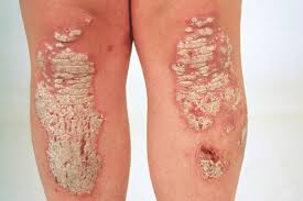
Psoriasis is a skin disorder that causes skin cells to multiply up to 10 times faster than normal. This makes the skin build up into bumpy red patches covered with white scales. They can grow anywhere, but most appear on the scalp, elbows, knees, and lower back. Psoriasis can't be passed from person to person. It does sometimes happen in members of the same family.
PSORIASIS  

PSORIASIS

 Psoriasis usually appears in early adulthood. For most people, it affects just a few areas. In severe cases, psoriasis can cover large parts of the body. The patches can heal and then come back throughout a person's life.get the basics on psoriasis
SYMPTOMS
The symptoms of psoriasis vary depending on the type you have. Some common symptoms for plaque psoriasis -- the most common variety of the condition -- include
Plaques of red skin, often covered with silver-colored scales. These plaques may be itchy and painful, and they sometimes crack and bleed. In severe cases, the plaques will grow and merge, covering large areas.
Disorders of the fingernails and toenails, including discoloration and pitting of the nails. The nails may also crumble or detach from the nail bed.
Plaques of scales or crust on the scalp.
People with psoriasis can also get a type of arthritis called psoriatic arthritis. It causes pain and swelling in the joints. The National Psoriasis Foundation estimates that between 10% to 30% of people with psoriasis also have psoriatic arthritis.
TYPES
Other types of psoriasis include:
PUSTULAR PSORIASIS- which causes red and scaly skin with tiny pustules on the palms of the hands and soles of the feet.
GUTTATE PSORIASIS- which often starts in childhood or young adulthood, causes small, red spots, mainly on the torso and limbs. Triggers may be respiratory infections, strep throat, tonsillitis, stress, injury to the skin, and taking antimalarial and beta-blocker medications.
INVERSE PSORIASIS- which makes bright red, shiny lesions that appear in skin folds, such as the armpits, groin, and under the breasts.
ERYTHRODERMIC PSORIASIS- which causes fiery redness of the skin and shedding of scales in sheets. It's triggered by severe sunburn, infections, certain medications, and stopping some kinds of psoriasis treatment. It needs to be treated immediately because it can lead to severe illness.
CAUSES
No one knows the exact cause of psoriasis, but experts believe that it’s a combination of things. Something wrong with the immune system causes inflammation, triggering new skin cells to form too quickly. Normally, skin cells are replaced every 10 to 30 days. With psoriasis, new cells grow every 3 to 4 days. The buildup of old cells being replaced by new ones creates those silver scales.
Psoriasis tends to run in families, but it may be skip generations. For instance, a grandfather and his grandson may be affected, but not the child's mother.
Things that can trigger an outbreak of psoriasis include:
  • Cuts, scrapes, or surgery
  • Emotional stress
  • Strep infections
  • Medications, including
  • Blood pressure medications (like beta-blockers)
  • Hydroxychloroquine, antimalarial medication
  • Diagnosis
  • Physical exam. It’s usually easy for your doctor to diagnose psoriasis, especially if you have plaques on areas such as your:
  • Scalp
  • Ears
  • Elbows
  • Knees
  • Belly button
  • Nails
Lab tests. The doctor might do a biopsy -- remove a small piece of skin and test it to make sure you don’t have a skin infection. There’s no other test to confirm or rule out psoriasis.
TREATMENT
Luckily, there are many treatments. Some slow the growth of new skin cells, and others relieve itching and dry skin. Your doctor will select a treatment plan that is right for you based on the size of your rash, where it is on your body, your age, your overall health, and other things. Common treatments include:
STEROIDS CREAM
Moisturizers for dry skin
Coal tar (a common treatment for scalp psoriasis available in lotions, creams, foams, shampoos, and bath solutions)
cream or ointment (a strong kind ordered by your doctor. Vitamin D in foods and pills has no effect.)
Retinoid creams
Treatments for moderate to severe psoriasis include:
LIGHT THERAPY- A doctor shines ultraviolet light on your skin to slow the growth of skin cells. PUVA is a treatment that combines a medicine called psoralen with a special form of ultraviolet light.
PSORIASISPSORIASIS LIGHT THERAPY
  PSORIASIS LIGHT THERAPY

 METHOTRAXATE- This drug can cause bone marrow and liver disease as well as lung problems, so it’s only for serious cases. Doctors closely watch patients. You will have to get lab tests, perhaps a chest X-ray, and possibly a liver biopsy.
RETINOIDS These pills, creams, foams, lotions, and gels are a class of drugs related to vitamin A. Retinoids can cause serious side effects, including birth defects, so they’re not recommended for women who are pregnant or planning to have children.
. This drug, made to suppress the immune system, may be taken for serious cases that do not respond to other treatments. It can damage the kidneys and raise blood pressure, so your doctor will closely watch your health while you take it.
BIOLOGIC TREATMENT-These work by blocking the body's immune system (which is overactive in psoriasis) to better control the inflammation from psoriasis. Biologic medications include adalimumab (Humira), brodalumab (Siliq), certolizumab pegol (Cimzia) etanercept (Enbrel), guselkumab (Tremfya), infliximab (Remicade), ixekizumab (Taltz), secukinumab (Cosentyx), tildrakizumab (Ilumya), and ustekinumab (Stelara).
An enzyme inhibitor. The medication apremilast (Otezla) is a new kind of drug for long-term inflammatory diseases like psoriasis and psoriatic arthritis. It's a pill that blocks a specific enzyme, which helps to slow other reactions that lead to inflammation.

PHOTOTHERAPY
Phototherapy or light therapy, involves exposing the skin to ultraviolet light on a regular basis and under medical supervision. Treatments are done in a doctor's office or psoriasis clinic or at home with phototherapy unit. The key to success with light therapy is consistency.
National Psoriasis Foundation does not support the use of indoor tanning beds as a substitute for phototherapy performed with a prescription and under a doctor's supervision. Indoor tanning raises the risk of melanoma by 59 percent, according to the American Academy of Dermatology and the World Health Organization, and does not provide the type of light that most effectively treats psoriasis. Read more on the Psoriasis Foundation position on indoor tanning beds »
Find a provider who offers phototherapy in our Health Care Provider Directory »
Learn more about phototherapy by contacting our Patient Navigation Center »
  • Learn about different types of light therapy.
    Ultraviolet light B (UVB)
  • Sunlight
  • Psoralen + UVA (PUVA)
  • Laser Treatments
  • Tanning beds
ULTRAVIOLET LIGHT B (UVB)
UVB PHOTOTHERAPY
Present in natural sunlight, ultraviolet B (UVB) is an effective treatment for psoriasis. UVB penetrates the skin and slows the growth of affected skin cells. Treatment involves exposing the skin to an artificial UVB light source for a set length of time on a regular schedule. This treatment is administered in a medical setting or at home.
There are two types of UVB treatment, broad band and narrow band. The major difference between them is that narrow band UVB light bulbs release a smaller range of ultraviolet light. Narrow-band UVB is similar to broad-band UVB in many ways. Several studies indicate that narrow-band UVB clears psoriasis faster and produces longer remissions than broad-band UVB. It also may be effective with fewer treatments per week than broad-band UVB.
During UVB treatment, your psoriasis may worsen temporarily before improving. The skin may redden and itch from exposure to the UVB light. To avoid further irritation, the amount of UVB administered may need to be reduced. Occasionally, temporary flares occur with low-level doses of UVB. These reactions tend to resolve with continued treatment.
UVB can be combined with other topical and/or systemic agents to enhance efficacy, but some of these may increase photosensitivity and burning, or shorten remission. Combining UVB with systemic therapies may increase efficacy dramatically and allow for lower doses of the systemic medication to be used.
UVB treatment is offered in different ways. This can include small units for localized areas such as the hands and feet, full body units or handheld units. Some UVB units use traditional UV lamps or bulbs, and others use LED bulbs.
HOME UVB PHOTOTHERAPY
Treating psoriasis with a UVB light unit at home is an economical and convenient choice for many people. Like phototherapy in a clinic, it requires a consistent treatment schedule. Individuals are treated initially at a medical facility and then begin using a light unit at home.
It is critical when doing phototherapy at home to follow a doctor's instructions and continue with regular check-ups. Home phototherapy is a medical treatment that requires monitoring by a health care professional.
All phototherapy treatments, including purchase of equipment for home use, require a prescription. Some insurance companies will cover the cost of home UVB equipment. Vendors of home phototherapy equipment often will assist you in working with your insurance company to purchase a unit.
SUNLIGHT
Although both UVB and ultraviolet light A (UVA) are found in sunlight, UVB works best for psoriasis. UVB from the sun works the same way as UVB in phototherapy treatments.
Short, multiple exposures to sunlight are recommended. Start with five to 10 minutes of noontime sun daily. Gradually increase exposure time by 30 seconds if the skin tolerates it. To get the most from the sun, all affected areas should receive equal and adequate exposure. Remember to wear sunscreen on areas of your skin unaffected by psoriasis.
Avoid overexposure and sunburn. It can take several weeks to see improvement. Have your doctor check you regularly for sun damage.
Some topical medications can increase the risk of sunburn. These include tazarotene, coal tar, Elidel (pimecrolimus) and Protopic (tacrolimus). Individuals using these products should talk with a doctor before going in the sun.
People who are using PUVA or other forms of light therapy should limit or avoid exposure to natural sunlight unless directed by a doctor.

PSORALEN + UVA (PUVA)
Like UVB, ultraviolet light A (UVA) is present in sunlight. Unlike UVB, UVA is relatively ineffective unless used with a light-sensitizing medication psoralen, which is administered topically or orally. This process, called PUVA, slows down excessive skin cell growth and can clear psoriasis symptoms for varying periods of time. Stable plaque psoriasis, guttate psoriasis, and psoriasis of the palms and soles are most responsive to PUVA treatment.
[caption id="attachment_1758" align="aligncenter" width="550"]PUVA PUVA[/caption]The most common short-term side effects of PUVA are nausea, itching and redness of the skin. Drinking milk or ginger ale, taking ginger supplements or eating while taking oral psoralen may prevent nausea. Antihistamines, baths with colloidal oatmeal products or application of topical products with capsaicin may help relieve itching. Swelling of the legs from standing during PUVA treatment may be relieved by wearing support hose.
LASER TRETMENT
EXCIMER LASER
The excimer laser—recently approved by the Food and Drug Administration (FDA) for treating chronic, localized psoriasis plaques—emits a high-intensity beam of ultraviolet light B (UVB).
The excimer laser can target select areas of the skin affected by mild to moderate psoriasis, and research indicates it is a particularly effective treatment for scalp psoriasis. Researchers at the University of Utah, for example, reported in The Journal of Drugs in Dermatology that in a small series of patients, laser treatment, combined with a topical steroid, cleared scalp psoriasis that resisted other treatment.

Individual response to the treatment varies. It can take an average of four to 10 sessions to see results, depending on the particular case of psoriasis. It is recommended that patients receive two treatments per week, with a minimum of 48 hours between treatments.
There is not yet enough long-term data to indicate how long the improvement will last following a course of laser therapy.

TANNING BEDS
Some people visit tanning salons as an alternative to natural sunlight. Tanning beds in commercial salons emit mostly UVA light, not UVB. The beneficial effect for psoriasis is attributed primarily to UVB light. National Psoriasis Foundation does not support the use of indoor tanning beds as a substitute for phototherapy performed with a prescription and under a doctor's supervision. Read more on the Psoriasis Foundation position on indoor tanning bed.

The American Academy of Dermatology, the Food and Drug Administration (FDA) and the Centers for Disease Control and Prevention all discourage the use of tanning beds and sun lamps. Indoor tanning raises the risk of melanoma by 59 percent, according to the American Academy of Dermatology and the World Health Organization. In May 2014, the FDA reclassified sunlamps (which are used in tanning beds and booths) from Class I (low risk) to Class II (moderate risk) products. The FDA can exert more regulatory control over Class II products, according to a press release on the FDA website.
The ultraviolet radiation from these devices can damage the skin, cause premature aging and increase the risk of skin cancer.

Wednesday 21 November 2018

TATROLOGY OF FALLOT

INTRODUCTION
Tetralogy of Fallot (teh-TRAL-uh-jee of fuh-LOW) is a rare condition caused by a combination of four heart defects that are present at birth (congenital).

These defects, which affect the structure of the heart, cause oxygen-poor blood to flow out of the heart and to the rest of the body. Infants and children with tetralogy of Fallot usually have blue-tinged skin because their blood doesn't carry enough oxygen.

Tetralogy of Fallot is often diagnosed during infancy or soon after. However, tetralogy of Fallot might not be detected until later in life in some adults, depending on the severity of the defects and symptoms.
With early diagnosis followed by appropriate surgical treatment, most children and adults who have tetralogy of Fallot live relatively normal lives, though they'll need regular medical care throughout life and might have restrictions on exercise
.
SYMPTOMS
Tetralogy of Fallot symptoms vary, depending on the extent of obstruction of blood flow out of the right ventricle and into the lungs. Signs and symptoms may include:
A bluish coloration of the skin caused by blood low in oxygen (cyanosis)
Shortness of breath and rapid breathing, especially during feeding or exercise
Loss of consciousness (fainting)
Clubbing of fingers and toes — an abnormal, rounded shape of the nail bed
Poor weight gain
Tiring easily during play or exercise
Irritability
Prolonged crying
A heart murmur
Tet spells
Sometimes, babies who have tetralogy of Fallot will suddenly develop deep blue skin, nails and lips after crying or feeding, or when agitated.
These episodes are called tet spells and are caused by a rapid drop in the amount of oxygen in the blood. Tet spells are most common in young infants, around 2 to 4 months old. Toddlers or older children might instinctively squat when they're short of breath. Squatting increases blood flow to the lungs.
Seek medical help if you notice that your baby has the following symptoms:
  • Difficulty breathing
  • Bluish discoloration of the skin
  • Passing out or seizures
  • Weakness
  • Unusual irritability
  • If your baby becomes blue (cyanotic), place your baby on his or her side and pull your baby's knees up to his or her chest. This helps increase blood flow to the lungs.
CAUSES
 
                                                       Tatrology of fallot
Tetralogy of Fallot occurs during fetal growth, when the baby's heart is developing. While factors such as poor maternal nutrition, viral illness or genetic disorders might increase the risk of this condition, in most cases the cause of tetralogy of Fallot is unknown.
The four abnormalities that make up the tetralogy of Fallot include:
  • PULMONARY VALVE STENOSIS- Pulmonary valve stenosis is a narrowing of the pulmonary valve — the valve that separates the lower right chamber of the heart (right ventricle) from the main blood vessel leading to the lungs (pulmonary artery).
Narrowing (constriction) of the pulmonary valve reduces blood flow to the lungs. The narrowing might also affect the muscle beneath the pulmonary valve. In some severe cases, the pulmonary valve doesn't form properly (pulmonary atresia) and causes reduced blood flow to the lungs.
  • VENTRICULAR SEPTAL DEFECT- A ventricular septal defect is a hole (defect) in the wall (septum) that separates the two lower chambers of the heart -the left and right ventricles. The hole allows deoxygenated blood in the right ventricle — blood that has circulated through the body and is returning to the lungs to replenish its oxygen supply — to flow into the left ventricle and mix with oxygenated blood fresh from the lungs.
Blood from the left ventricle also flows back to the right ventricle in an inefficient manner. This ability for blood to flow through the ventricular septal defect reduces the supply of oxygenated blood to the body and eventually can weaken the heart.
  • OVERRIDING AORTA- Normally the aorta — the main artery leading out to the body — branches off the left ventricle. In tetralogy of Fallot, the aorta is shifted slightly to the right and lies directly above the ventricular septal defect.
In this position the aorta receives blood from both the right and left ventricles, mixing the oxygen-poor blood from the right ventricle with the oxygen-rich blood from the left ventricle.
  • RIGHT VENTRICULAR HYPERTROPHY- When the heart's pumping action is overworked, it causes the muscular wall of the right ventricle to thicken. Over time this might cause the heart to stiffen, become weak and eventually fail.
    Some children or adults who have tetralogy of Fallot may have other heart defects, such as a hole between the heart's upper chambers (atrial septal defect), a right aortic arch or abnormalities of the coronary arteries.
RISK FACTOR
While the exact cause of tetralogy of Fallot is unknown, various factors might increase the risk of a baby being born with this condition. These risk factors include:
  • A viral illness during pregnancy, such as rubella (German measles)
  • Alcoholism during pregnancy
  • Poor nutrition during pregnancy
  • A mother older than age 40
  • A parent who has tetralogy of Fallot
  • The presence of Down syndrome or DiGeorge syndrome
COMPLICATIONS
All babies who have tetralogy of Fallot need corrective surgery. Without treatment, your baby might not grow and develop properly.
Your baby may also be at an increased risk of serious complications, such as infective endocarditis — an infection of the inner lining of the heart or heart valve caused by a bacterial infection.
Untreated cases of tetralogy of Fallot usually develop severe complications over time, which might result in death or disability by early adulthood.

PHYSICAL THERAPY
 
PRE-OPERATIVE – Most infants with TOF will receive thoracic surgery to correct the associated abnormalities. The most important physical therapy intervention pre-operative for the patient and family is education. Many institutes are using a method of education that describes a pre-operative program that helps to decrease post-operative complications. Many institutions use a doll to explain to children about the tubes and how to care for them.
POST-OPERATIVE– Pulmonary function is a main area of concentration after thoracic surgery. The interventions vary with age but the general goal is the same, to mobilize secretions, increase aeration and increase general mobility. Atelectasis is a concern after thoracic surgery and is caused by the slowed mucous transport, altered breathing patterns, prolonged positioning in supine and diaphragmatic dysfunction in early post-operative period. The yawn maneuver and prolonged inspiration to increase inflation are ways to prevent atelectasis. The incentive spirometry is an effective tool for preventing atelectasis in the pediatric population. Other ways that can be more fun for children are activities such as blowing bubbles or blowing on a windmill. There are expiratory maneuvers but the children typically take a large breath before blowing, thus they become inspiratory maneuvers also.

Segmental expansion techniques are effective to increase segmental aeration. These techniques are performed by placing your hand over whichever segment of the lung is not effectively inflating and allowing your hand to move with the respiratory cycle. Gentle pressure may be applied to the chest at the end of the expiratory phase, just before the inhalation phase. This elicits a stretch reflex that facilitates air flow to that particular segment.

Percussion and vibration may be performed in conjunction with segmental expansion. Percussion is the rhythmic clapping with cupped hands over the involved lung segment performed throughout the respiratory cycle, with the goal of mechanically dislodging pulmonary secretions. Vibration is performed by creating a fine oscillating movement of the hands on the chest wall just before expiration begins and throughout the expiration phase. If the child does not tolerate percussion and vibration, placing the patient in side lying and gently rocking back and forth may stimulate expansion, secretion motion and relaxation. This technique is affective if the child is upset and may decrease respiratory rate.

Positioning may be useful for patients, but certain positions may be contraindicated post thoracic surgery. Check with the MD or nurse before using positioning to make sure that it is safe for the child.

POSTURAL DRAINAGE
If the child is intubated suctioning may be used to remove secretions from the airways, however children with cyanotic defects (such as TOF) tend to desaturate during suctioning and it is very important to hyperventilate these patients before and after suctioning and to monitor oxygen levels.
Coughing is an effective way to mobilize secretions if the patient is able and willing to cough. A teddy bear or pillow can be used by the child to self-splint the incision. Have the child squeeze the stuffed animal against their chest to decrease pain.

Range of motion (ROM) exercises should be initiated as soon as possible after surgery. ROM exercises are extremely important with thoracic surgery because this type of incision tends to produce more guarding. Child position should be changed regularly to avoid a pooling of secretions in the dependent part of the lungs and regular position changes reduce the risk of fevers after surgery. Ambulation should be implemented as soon as possible to decrease both pulmonary and circulatory complications. As soon as atrial lines and groin lines are removed, patients should begin to ambulate, even if only 5-10 ft.

DIFFERENTIAL DIAGNOSIS
Any other cyanotic heart defects, such as, pulmonary stenosis, transposed arterial trunks, common arterial trunk and tricuspid atresia. Patients with heart murmurs will also be tests for persistent pulmonary hypertension.